A clinical reference for those who examine tooth wear chairside
Tooth wear: what it is, its three mechanisms, and when it stops being physiological
Dr. Guilherme Cabral
Tooth wear is the cumulative surface loss of mineralized tooth substance due to physical or chemo-physical processes not related to dental caries [1].
1. FDI World Dental Federation. Tooth wear. FDI policy statement. Int Dent J. 2024;74(1):163-164. PMID 38218598. PMCID PMC10829352. DOI 10.1016/j.identj.2023.10.007.
Enamel does not grow back, so every loss adds up over a lifetime. That is why some wear comes with age, and that much is physiological. In the chair, the question is whether a patient has lost more than their age explains. If they have, the next question is which mechanism is behind it.
Types of tooth wear: biocorrosion (erosion), tension (abfraction), and friction (abrasion, attrition)
Here, the four names fit into three mechanisms: acid dissolves tissue, load flexes the tooth, contact wears the surface. Each tends to leave its own shape on the tooth, and that shape is the first clue you read.
Biocorrosion (erosion)
What it is
Biocorrosion (erosion) is mineral loss by chemical dissolution. The acid comes from the diet or from the stomach. Grippo and colleagues proposed the term biocorrosion for this chemical action [2].
2. Grippo JO, Simring M, Coleman TA. Abfraction, abrasion, biocorrosion, and the enigma of noncarious cervical lesions: a 20-year perspective. J Esthet Restor Dent. 2012;24(1):10-23. PMID 22296690. DOI 10.1111/j.1708-8240.2011.00487.x.
How it happens
Hydrogen ions from the acid break down hydroxyapatite and release calcium into the saliva, on every surface they reach, with no preferred direction. Before any loss is visible, the surface softens. Saliva can still reharden that layer if nothing rubs it away; what has already dissolved does not come back.
How it presents
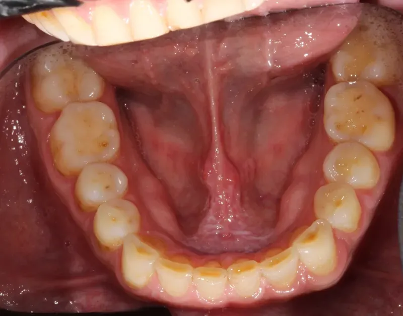
Dissolution tends to leave a rounded outline, wider than it is deep. The enamel surface turns smooth and glossy. On posterior teeth, the cusps round off and the occlusal anatomy fades.
In the cervical region, the attack stops just short of the gingival margin and often leaves a thin band of intact enamel standing. Which surface is involved suggests the source: on the facial of anterior teeth, dietary acid; on the palatal of maxillary anterior teeth, gastric acid.
These are the usual sites, not the only ones.
Tension (abfraction)
What it is
Tension (abfraction) is the share of cervical loss that comes from load. When load bends the tooth, tensile stress concentrates in the cervical region and helps break down enamel and dentin.
How it happens
Load along the long axis becomes compression, which the tooth tolerates well. Oblique load bends the tooth like a cantilever anchored in bone, and tensile stress concentrates in the cervical third. The enamel there is thin, and enamel resists tension far less than compression. A single load leaves no mark; repeated, the load weakens the tissue, more so where acid has softened it. Occlusion decides where the stress lands, and a clenching or grinding habit decides the dose.
How it presents
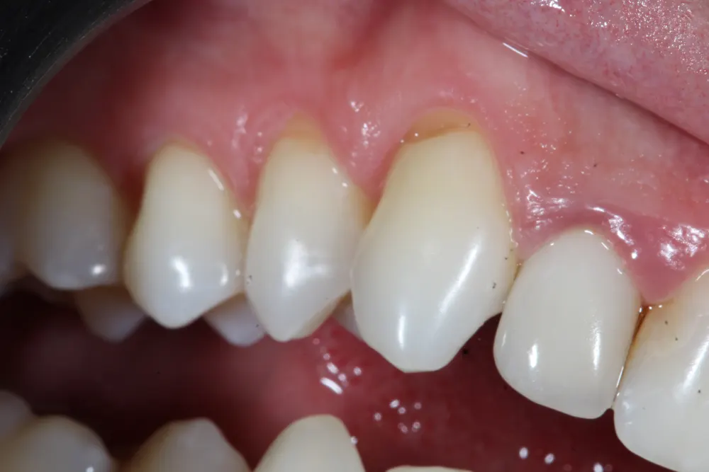
The lesion starts on the surface that flexure stretches, which depends on the cuspal incline taking the load. The shape tends toward a wedge, with a sharp internal angle, flat walls, and a straight margin; acid and friction are often at work in that same wedge. With a thick gingival phenotype, the lesion can grow below the margin, and there clenching and grinding are among the first questions. With a thin phenotype, the same load can make the bone resorb and the margin recede, which leaves the exposed root within reach of the other mechanisms.
These are the usual sites, not the only ones.
Friction (abrasion, attrition)
What it is
Friction (abrasion, attrition) is loss from mechanical contact. There are two types, told apart by what touches the tooth.
Where friction weighs in
On tissue that acid has softened or load has flexed, contact speeds up the loss. In vitro, acid-softened enamel lost four times more to toothbrushing than sound enamel [3].
3. Wiegand A, Köwing L, Attin T. Impact of brushing force on abrasion of acid-softened and sound enamel. Arch Oral Biol. 2007;52(11):1043-7. PMID 17658454. DOI 10.1016/j.archoralbio.2007.06.004.
Attrition: tooth against tooth
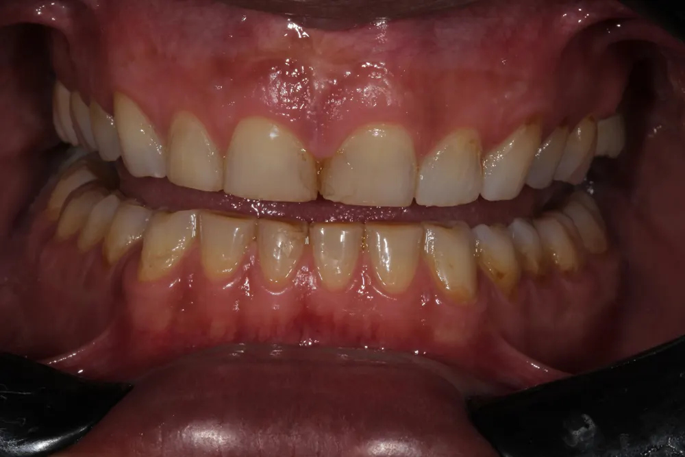
In attrition, with nothing between the teeth, repeated contact flattens the spot where they touch. It comes from bruxism, during sleep or while awake. It usually shows where teeth meet: occlusal surfaces, incisal edges, cusp tips, and the tips of the canines. The mark is a flat, polished facet. In closure and excursions, the facet on one tooth fits the facet on its antagonist.
Abrasion: an outside agent against the tooth
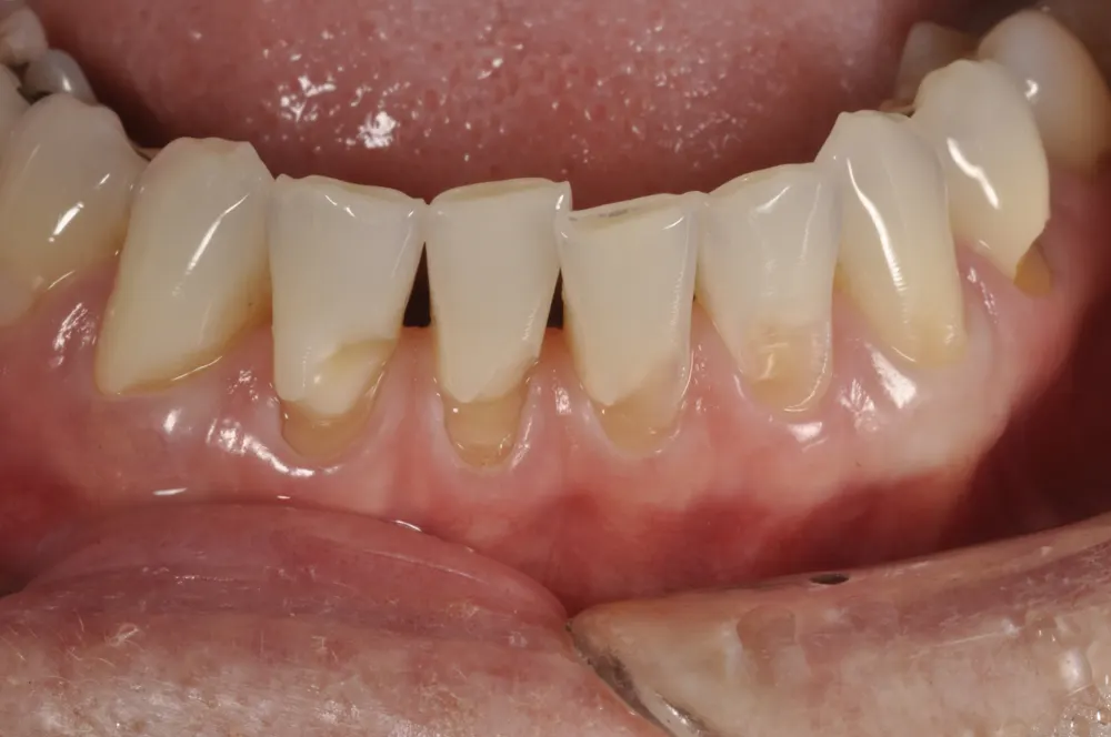
In abrasion from toothbrushing, the particles in the toothpaste do the cutting; the brush only carries them. Fibrous food and objects held in the mouth abrade the tooth too. Abrasion usually shows on the facial surface and in the cervical region, along the path of the brush, as shallow, crisscrossing scratches on a rough, matte surface. On sound enamel, the brush removes little. Its direct damage is greater on the gingiva, which recedes and exposes the root.
These are the usual sites, not the only ones.
| Mechanism | What acts | Where it usually shows | What it looks like |
|---|---|---|---|
| Biocorrosion (erosion) | Acid from the diet or the stomach, not from bacteria, that dissolves tissue | Facial of anterior teeth (dietary acid), palatal of maxillary anterior teeth (gastric acid), occlusal of posterior teeth | Tends toward a rounded outline, wider than it is deep; on enamel, a smooth, glossy surface; cervically, often a thin band of intact enamel at the gingival margin |
| Tension (abfraction) | Oblique load that bends the tooth; the tensile stress from that flexure helps break down enamel and dentin | Cervical third, on the surface that flexure stretches; with a thick gingival phenotype, it can grow below the margin | Tends toward a wedge: sharp internal angle, flat walls, straight margin; often with acid and friction at work too |
| Friction (abrasion, attrition) | Attrition: tooth against tooth, from bruxism. Abrasion: an outside agent (toothpaste particles, fibrous food, objects) | Attrition: where teeth meet (occlusal surfaces, incisal edges, cusp tips). Abrasion: facial and cervical surfaces, along the path of the brush | Attrition: a flat, polished facet that fits the one on its antagonist. Abrasion: shallow, crisscrossing scratches on a rough, matte surface |
These are the usual sites, not the only ones.
One lesion, more than one mechanism
A non-carious cervical lesion (NCCL) is a loss of tooth structure in the cervical region from causes other than caries. Teixeira and colleagues' 2020 systematic review of 24 studies put the weighted mean prevalence of NCCLs in adults at 46.7% [4]. Goodacre and colleagues describe NCCLs as multifactorial in etiology [5]. Take a patient who sucks on oranges through the day. The citric acid softens the cervical surface, and the fiber of the fruit, chew after chew, carries away what the acid softened. Two mechanisms, one lesion. The same can happen on a cusp, which hollows into a cup ringed by enamel. An in vitro study by Ruben and colleagues produced cups only when acid at pH 4.8 was combined with a load of 30 or 50 N; at pH 5.5, or with no load, none formed [6]. A cupped cusp sends you to ask about acid and about load. Which one leads changes from patient to patient: in one it is load, in the next it is acid.
4. Teixeira DNR, Thomas RZ, Soares PV, Cune MS, Gresnigt MMM, Slot DE. Prevalence of noncarious cervical lesions among adults: a systematic review. J Dent. 2020;95:103285. PMID 32006668. DOI 10.1016/j.jdent.2020.103285.
5. Goodacre CJ, Roberts WE, Munoz CA. Noncarious cervical lesions: morphology and progression, prevalence, etiology, pathophysiology, and clinical guidelines for restoration. J Prosthodont. 2023;32(2):e1-e18. PMID 35920595. DOI 10.1111/jopr.13585.
6. Ruben JL, Roeters FJM, Truin GJ, Loomans BAC, Huysmans MDNJM. Cup-shaped tooth wear defects: more than erosive challenges? Caries Res. 2019;53(4):467-474. PMID 30840963. PMCID PMC7050669. DOI 10.1159/000496983.
Within one lesion, read it wall by wall. Is the outline rounded, or does it narrow into a wedge? Look at where the walls meet, and note whether the angle is sharp or soft. Then check the surface inside the concavity for scratches.
The shape raises a suspicion about the mechanism. The consultation confirms it: the history you take, and the reasoning you work through with the patient.
BEWE: the index that grades the extent of loss
The Basic Erosive Wear Examination (BEWE) scores each surface from 0 to 3, first by the texture of the surface and then by the fraction of its area lost. It records the highest score in each sextant, and the sum of the six sextants guides management [7]. Two surfaces with the same area lost get the same score, whatever the mechanism and whatever the rate of loss.
7. Bartlett D, Ganss C, Lussi A. Basic Erosive Wear Examination (BEWE): a new scoring system for scientific and clinical needs. Clin Oral Investig. 2008;12(Suppl 1):S65-S68. PMID 18228057. PMCID PMC2238785. DOI 10.1007/s00784-007-0181-5.
Tap a score for its criterion.
No erosive tooth wear. The surface is intact.
Initial loss of surface texture. There is no defect with a defined outline yet: what has changed is the texture.
Distinct defect: hard tissue loss of less than 50% of the surface area. Dentin is often involved, but area sets the score, not dentin.
Hard tissue loss of 50% or more of the surface area. Here too dentin is often involved, and the score still comes from area.
In 2026, Bartlett, Lussi and colleagues proposed BEWE 2.0, which adds a score of 4 for 2 mm of lost crown height or 2 mm of horizontal wear along the cervical margin and clarifies the line between scores 0 and 1 [8]. They describe the original index as a simple screening tool. The risk levels in the table below come from the 2008 version.
8. Bartlett D, O'Toole S, Chen V, et al. BEWE 2.0: Basic erosive tooth wear examination revisited: introducing an additional level for more severe erosive tooth wear. J Dent. 2026;166:106514. PMID 41565150. DOI 10.1016/j.jdent.2026.106514.
| Sum of the six sextants | BEWE risk level (2008) |
|---|---|
| 2 or less | None |
| 3 to 8 | Low |
| 9 to 13 | Medium |
| 14 or more | High |
From Bartlett, Ganss and Lussi, 2008. The authors note that the cut-off values come from one author's experience and studies, and that they have to be reconsidered.
The criteria name no agent, though the index was built for erosive wear. The management the 2008 paper ties to each risk level leans toward acid, with diet and fluoride [7]; when the loss comes from load, the score still records it, and that advice misses part of the cause. A risk level also counts only the loss already there; exposure that has not marked the tooth yet is read in the patient.
7. Bartlett D, Ganss C, Lussi A. Basic Erosive Wear Examination (BEWE): a new scoring system for scientific and clinical needs. Clin Oral Investig. 2008;12(Suppl 1):S65-S68. PMID 18228057. PMCID PMC2238785. DOI 10.1007/s00784-007-0181-5.
Time and the person separate physiological from pathological wear
Loomans and colleagues, in the European consensus on severe tooth wear, pose the deciding question: is the wear consistent with the patient's age, or atypical for it [9]?
9. Loomans B, Opdam N, Attin T, et al. Severe tooth wear: European consensus statement on management guidelines. J Adhes Dent. 2017;19(2):111-119. PMID 28439579. DOI 10.3290/j.jad.a38102.
Take two patients with the same loss of structure on the same surface. One is 60, the other 24. At 60, decades of function can account for that loss, and the wear is consistent with age, unless an active cause turns up. At 24, the same loss built up in less than half the time: the wear has advanced faster than age explains, and that is where it reads as pathological.
Consistent with age: physiological
Atypical for age: pathological
Frequently asked questions
What causes tooth wear?
Unlike caries, tooth wear has no single agent such as bacteria: it is shaped by the patient's habits, lifestyle and health. Among 3,187 adults aged 18 to 35 in seven European countries, Bartlett and colleagues found wear associated with heartburn or acid reflux, repeated vomiting, snoring, and fresh fruit and juice [10]. None of these can be confirmed from the tooth alone; they come out in the history. To see how The Tooth Wear Workup teaches that history, visit the program page.
10. Bartlett DW, Lussi A, West NX, Bouchard P, Sanz M, Bourgeois D. Prevalence of tooth wear on buccal and lingual surfaces and possible risk factors in young European adults. J Dent. 2013;41(11):1007-13. PMID 24004965. DOI 10.1016/j.jdent.2013.08.018.
What should be done about tooth wear lesions? Isn't restoring them enough?
Restoring is one part of managing tooth wear lesions, and usually a late one. The European consensus guidelines on severe tooth wear recommend diagnosis, prevention, counseling and monitoring, to clarify the cause of the wear and how to control it. They advise that restorative intervention is typically best delayed as long as possible [9]. When a restoration is indicated, the same guidelines favor a conservative, minimally invasive approach, backed by preventive measures.
9. Loomans B, Opdam N, Attin T, et al. Severe tooth wear: European consensus statement on management guidelines. J Adhes Dent. 2017;19(2):111-119. PMID 28439579. DOI 10.3290/j.jad.a38102.
When does a non-carious cervical lesion need a restoration?
When comparable records show it progressing, even without pain; when sensitivity does not settle; when the loss compromises function; or when the patient has an esthetic demand. Goodacre and colleagues recommend prevention first and delaying restoration until it becomes necessary; among the factors they cite are lesion progression, sensitivity and poor esthetics [5]. Whichever trigger applies, find the cause first and place the restoration alongside its control or after it; a restoration gives back form and function and leaves the cause untouched.
5. Goodacre CJ, Roberts WE, Munoz CA. Noncarious cervical lesions: morphology and progression, prevalence, etiology, pathophysiology, and clinical guidelines for restoration. J Prosthodont. 2023;32(2):e1-e18. PMID 35920595. DOI 10.1111/jopr.13585.
Aren't non-carious cervical lesions caused by occlusion?
Occlusion can add to a non-carious cervical lesion (NCCL), but on its own it rarely explains one. A systematic review by Duangthip and colleagues covered 69 studies, 38 laboratory and 31 clinical. Across all 69, 81% reported an association between occlusal stress and NCCLs, yet no clinical study showed a lesion caused by stress alone [11]. In the mouth, load works alongside acid and contact and the loss comes from the combination. An occlusal finding opens the history; it does not close it.
11. Duangthip D, Man A, Poon PH, Lo ECM, Chu CH. Occlusal stress is involved in the formation of non-carious cervical lesions. A systematic review of abfraction. Am J Dent. 2017;30(4):212-220. PMID 29178704.
Isn't cervical wear just from brushing too hard?
No single brushing factor, hard brushing included, has been established as the primary cause of cervical abrasion. Reviewing the evidence, Litonjua and colleagues found the studies on force, technique, bristle stiffness and toothpaste too limited and conflicting to single one out, and concluded that brushing may act together with biocorrosion (erosion) and, possibly, occlusal load [12]. Hard brushing still belongs in the history, alongside the questions about acid and load.
12. Litonjua LA, Andreana S, Cohen RE. Toothbrush abrasions and noncarious cervical lesions: evolving concepts. Compend Contin Educ Dent. 2005;26(11):767-8, 770-4, 776 passim. PMID 16300231.
Isn't a healthy diet good for the teeth?
A diet that is healthy for the body can still be hard on enamel and dentin. In a systematic review, Smits and colleagues found the vegetarian diet associated with higher odds of biocorrosion (erosion) (odds ratio 2.40), though most included studies were of poor quality [13]. One likely reason is that fruit and vegetables eaten across many meals and snacks bring acid to the teeth more often. The risk follows the eating pattern more than any single food.
13. Smits KPJ, Listl S, Jevdjevic M. Vegetarian diet and its possible influence on dental health: a systematic literature review. Community Dent Oral Epidemiol. 2020;48(1):7-13. PMID 31571246. PMCID PMC6972589. DOI 10.1111/cdoe.12498.
Isn't biocorrosion (erosion) about what the patient drinks?
Biocorrosion (erosion) is not only about what the patient drinks. Acid can also come from within, in reflux or repeated vomiting, and both are risk factors for tooth wear [10]. When the acid does come from a drink, how they drink it matters too. Johansson and colleagues compared young men with high and low levels of biocorrosion: the high group kept a cola-type drink in the mouth for 71 seconds, against 40, while the pH after drinking did not differ between the groups [14]. The study was small, 19 men in all, but it adds a question to the history: how each drink is taken.
10. Bartlett DW, Lussi A, West NX, Bouchard P, Sanz M, Bourgeois D. Prevalence of tooth wear on buccal and lingual surfaces and possible risk factors in young European adults. J Dent. 2013;41(11):1007-13. PMID 24004965. DOI 10.1016/j.jdent.2013.08.018.
14. Johansson AK, Lingström P, Birkhed D. Comparison of factors potentially related to the occurrence of dental erosion in high- and low-erosion groups. Eur J Oral Sci. 2002;110(3):204-11. PMID 12120705. DOI 10.1034/j.1600-0447.2002.11211.x.
Can tooth wear point to acid reflux?
It can raise the question; a physician answers it. In a 2020 meta-analysis, Jordão and colleagues found that people with gastroesophageal reflux disease, or its symptoms, had 2 to 4 times the odds of erosive tooth wear, though the studies varied widely [15]. The association was stronger when reflux was confirmed by testing (odds ratio 4.13) than when it rested on reported symptoms (2.69). Those odds measure who has the wear; progression was not part of the review. Smooth loss on the palatal of the maxillary anterior teeth, with the mandibular anterior teeth spared early on, points to acid from within. If diet and drinks do not explain it, you record it and refer, even when the patient reports no heartburn.
15. Jordão HWT, Coleman HG, Kunzmann AT, McKenna G. The association between erosive toothwear and gastro-oesophageal reflux-related symptoms and disease: a systematic review and meta-analysis. J Dent. 2020;95:103284. PMID 32006670. DOI 10.1016/j.jdent.2020.103284.
How do you treat a tooth wear lesion?
You treat a tooth wear lesion through the host first: the patient, and what their routine keeps doing to the tooth. The lesion is the part of the disease that has already happened, so the working rule is to control the host, not the lesion. For biocorrosion (erosion), the European Federation of Conservative Dentistry includes screening for early signs and evaluating all etiological factors in effective management [16].
16. Carvalho TS, Colon P, Ganss C, et al. Consensus report of the European Federation of Conservative Dentistry: erosive tooth wear diagnosis and management. Swiss Dent J. 2016;126(4):342-346. PMID 27142130. DOI 10.61872/sdj-2016-04-143.
Doesn't a night guard stop bruxism?
A night guard can protect the teeth during bruxism; the evidence does not show that it stops the bruxism itself. A Cochrane review by Macedo and colleagues found insufficient evidence that occlusal splints treat sleep bruxism, and noted that they may bring some benefit for tooth wear [17]. Ferreira and colleagues reviewed 12 studies in a 2024 systematic review and meta-analysis and found that occlusal appliances did not change masseter and temporalis activity or bite force in people with sleep bruxism. They rated the certainty of that evidence low to very low [18]. The muscles may keep working as hard as before, with the acrylic wearing in place of the enamel. So the splint can protect the teeth while you look for what drives the grinding.
17. Macedo CR, Silva AB, Machado MA, Saconato H, Prado GF. Occlusal splints for treating sleep bruxism (tooth grinding). Cochrane Database Syst Rev. 2007;(4):CD005514. PMID 17943862. PMCID PMC8890597. DOI 10.1002/14651858.CD005514.pub2.
18. Ferreira GF, Carletti TM, Gama LT, Magno MB, Maia LC, Rodrigues Garcia RCM. Influence of occlusal appliances on the masticatory muscle function in individuals with sleep bruxism: a systematic review and meta-analysis. Eur J Oral Sci. 2024;132(2):e12979. PMID 38421263. DOI 10.1111/eos.12979.
Why is exposed dentin sensitive in one patient and not in another?
Exposed dentin becomes sensitive when its tubules are open at the surface and reach the pulp, and not every worn surface has open tubules. Absi and colleagues compared exposed cervical dentin under the electron microscope: sensitive teeth had about eight times more tubules per area, about twice as wide, as non-sensitive teeth [19]. Addy and Pearce consider biocorrosion (erosion), which opens tubules, the probable main initiator of dentin hypersensitivity [20].
19. Absi EG, Addy M, Adams D. Dentine hypersensitivity. A study of the patency of dentinal tubules in sensitive and non-sensitive cervical dentine. J Clin Periodontol. 1987;14(5):280-4. PMID 3475295. DOI 10.1111/j.1600-051x.1987.tb01533.x.
20. Addy M, Pearce N. Aetiological, predisposing and environmental factors in dentine hypersensitivity. Arch Oral Biol. 1994;39(Suppl):33S-38S. PMID 7702465. DOI 10.1016/0003-9969(94)90186-4.
Is abfraction accepted?
Tension (abfraction) is proposed as one factor in non-carious cervical lesions, and debated as an explanation for them. Goodacre and colleagues note that publications have both supported and challenged the concept of abfraction [5]. Of the studies Duangthip and colleagues reviewed, 24 used finite element analysis and found stress concentrating in the cervical region [11]. A model shows where stress lands, not which lesion it made. In the chair, a wedge with a sharp internal angle is a clue to tension, not proof of it, and the next questions ask whether the patient clenches, grinds or brushes hard.
5. Goodacre CJ, Roberts WE, Munoz CA. Noncarious cervical lesions: morphology and progression, prevalence, etiology, pathophysiology, and clinical guidelines for restoration. J Prosthodont. 2023;32(2):e1-e18. PMID 35920595. DOI 10.1111/jopr.13585.
11. Duangthip D, Man A, Poon PH, Lo ECM, Chu CH. Occlusal stress is involved in the formation of non-carious cervical lesions. A systematic review of abfraction. Am J Dent. 2017;30(4):212-220. PMID 29178704.
Managing tooth wear: what to do, and in what order
Across all three mechanisms, management follows the same order. It starts with the person, and the patient carries out much of it.
Tap a step to read it
Start with the person
Before you touch a tooth, ask about their routine, diet, reflux, sleep, stress and medication, and take dated photographs. The shape of the wear tells you what to ask; only the patient can answer it.
Let the patient see it
Show them their own photographs and where the wear is heading if nothing changes. Then they decide how far to take treatment.
Control the cause, with the patient
The patient changes what is theirs to change; the rest goes to another clinician, with the dated photographs, as part of the treatment.
With acid, frequency is what counts: they space out the exposures, and right after one they rinse with water instead of brushing. Acid from within is not yours to treat: it gets referred.
With load, the patient learns to notice daytime clenching, and anxiety or poor sleep is referred. Canine guidance takes lateral load off the posterior teeth. Subtractive occlusal adjustment to treat the lesion is not supported by the evidence.
With contact, the patient changes how and when they brush, never how much: a soft brush and a low-abrasivity toothpaste.
Protect the tooth in the meantime
A hard stabilization splint takes the contact instead of the enamel and spares the surfaces that meet. If the history pointed to sleep apnea or night reflux, have that assessed first. Acute pain, an impending fracture or an exposed pulp is treated at once, alongside cause control. When the cause does not give way, protection becomes the strategy.
Restore last
A restoration gives back form and distributes load, but it does not treat the cause. Place it to give back function and structure, alongside cause control or after it.
Follow up without discharge
At each recall, photograph the same surface, compare it with the first record and revisit the habits with them. Keep the records comparable: the same framing and light for each photograph and, if you scanned or took an impression at the first visit, a new one of the same teeth. One record shows how much is gone. To know whether the wear is still active, you need a second one, comparable with the first. If the cause you targeted is under control and the wear has not slowed, the reading was wrong: reorder the plan.
The protocol for each measure, and the cases where a measure backfires, are taught in depth in The Tooth Wear Workup. One example: in a patient whose gastric acid rises at night, the bruxism splint holds that acid against the enamel.
See the programWe do not treat only the wear. We treat the person it happened to.
Start with the next case of tooth wear in your chair. Before you commit to any procedure, find the real cause of the wear, so you are not putting your work on the line.
Dr. Guilherme Cabral
References
- FDI World Dental Federation. Tooth wear. FDI policy statement. Int Dent J. 2024;74(1):163-164. PMID 38218598. PMCID PMC10829352. DOI 10.1016/j.identj.2023.10.007.
- Grippo JO, Simring M, Coleman TA. Abfraction, abrasion, biocorrosion, and the enigma of noncarious cervical lesions: a 20-year perspective. J Esthet Restor Dent. 2012;24(1):10-23. PMID 22296690. DOI 10.1111/j.1708-8240.2011.00487.x.
- Wiegand A, Köwing L, Attin T. Impact of brushing force on abrasion of acid-softened and sound enamel. Arch Oral Biol. 2007;52(11):1043-7. PMID 17658454. DOI 10.1016/j.archoralbio.2007.06.004.
- Teixeira DNR, Thomas RZ, Soares PV, Cune MS, Gresnigt MMM, Slot DE. Prevalence of noncarious cervical lesions among adults: a systematic review. J Dent. 2020;95:103285. PMID 32006668. DOI 10.1016/j.jdent.2020.103285.
- Goodacre CJ, Roberts WE, Munoz CA. Noncarious cervical lesions: morphology and progression, prevalence, etiology, pathophysiology, and clinical guidelines for restoration. J Prosthodont. 2023;32(2):e1-e18. PMID 35920595. DOI 10.1111/jopr.13585.
- Ruben JL, Roeters FJM, Truin GJ, Loomans BAC, Huysmans MDNJM. Cup-shaped tooth wear defects: more than erosive challenges? Caries Res. 2019;53(4):467-474. PMID 30840963. PMCID PMC7050669. DOI 10.1159/000496983.
- Bartlett D, Ganss C, Lussi A. Basic Erosive Wear Examination (BEWE): a new scoring system for scientific and clinical needs. Clin Oral Investig. 2008;12(Suppl 1):S65-S68. PMID 18228057. PMCID PMC2238785. DOI 10.1007/s00784-007-0181-5.
- Bartlett D, O'Toole S, Chen V, et al. BEWE 2.0: Basic erosive tooth wear examination revisited: introducing an additional level for more severe erosive tooth wear. J Dent. 2026;166:106514. PMID 41565150. DOI 10.1016/j.jdent.2026.106514.
- Loomans B, Opdam N, Attin T, et al. Severe tooth wear: European consensus statement on management guidelines. J Adhes Dent. 2017;19(2):111-119. PMID 28439579. DOI 10.3290/j.jad.a38102.
- Bartlett DW, Lussi A, West NX, Bouchard P, Sanz M, Bourgeois D. Prevalence of tooth wear on buccal and lingual surfaces and possible risk factors in young European adults. J Dent. 2013;41(11):1007-13. PMID 24004965. DOI 10.1016/j.jdent.2013.08.018.
- Duangthip D, Man A, Poon PH, Lo ECM, Chu CH. Occlusal stress is involved in the formation of non-carious cervical lesions. A systematic review of abfraction. Am J Dent. 2017;30(4):212-220. PMID 29178704.
- Litonjua LA, Andreana S, Cohen RE. Toothbrush abrasions and noncarious cervical lesions: evolving concepts. Compend Contin Educ Dent. 2005;26(11):767-8, 770-4, 776 passim. PMID 16300231.
- Smits KPJ, Listl S, Jevdjevic M. Vegetarian diet and its possible influence on dental health: a systematic literature review. Community Dent Oral Epidemiol. 2020;48(1):7-13. PMID 31571246. PMCID PMC6972589. DOI 10.1111/cdoe.12498.
- Johansson AK, Lingström P, Birkhed D. Comparison of factors potentially related to the occurrence of dental erosion in high- and low-erosion groups. Eur J Oral Sci. 2002;110(3):204-11. PMID 12120705. DOI 10.1034/j.1600-0447.2002.11211.x.
- Jordão HWT, Coleman HG, Kunzmann AT, McKenna G. The association between erosive toothwear and gastro-oesophageal reflux-related symptoms and disease: a systematic review and meta-analysis. J Dent. 2020;95:103284. PMID 32006670. DOI 10.1016/j.jdent.2020.103284.
- Carvalho TS, Colon P, Ganss C, et al. Consensus report of the European Federation of Conservative Dentistry: erosive tooth wear diagnosis and management. Swiss Dent J. 2016;126(4):342-346. PMID 27142130. DOI 10.61872/sdj-2016-04-143.
- Macedo CR, Silva AB, Machado MA, Saconato H, Prado GF. Occlusal splints for treating sleep bruxism (tooth grinding). Cochrane Database Syst Rev. 2007;(4):CD005514. PMID 17943862. PMCID PMC8890597. DOI 10.1002/14651858.CD005514.pub2.
- Ferreira GF, Carletti TM, Gama LT, Magno MB, Maia LC, Rodrigues Garcia RCM. Influence of occlusal appliances on the masticatory muscle function in individuals with sleep bruxism: a systematic review and meta-analysis. Eur J Oral Sci. 2024;132(2):e12979. PMID 38421263. DOI 10.1111/eos.12979.
- Absi EG, Addy M, Adams D. Dentine hypersensitivity. A study of the patency of dentinal tubules in sensitive and non-sensitive cervical dentine. J Clin Periodontol. 1987;14(5):280-4. PMID 3475295. DOI 10.1111/j.1600-051x.1987.tb01533.x.
- Addy M, Pearce N. Aetiological, predisposing and environmental factors in dentine hypersensitivity. Arch Oral Biol. 1994;39(Suppl):33S-38S. PMID 7702465. DOI 10.1016/0003-9969(94)90186-4.
Open-access papers on tooth wear are in the Files tab of the Toothwear Waiting Room (opens in a new tab), an open Facebook group for clinicians.